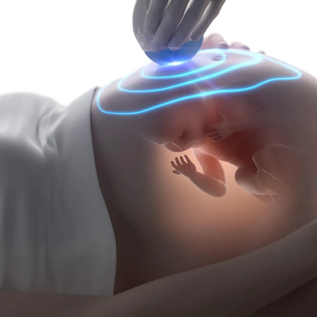
3RD TRIMESTER - GROWTH SCAN
Book an appointment
Discuss your case with the physician and find out what the next steps are.
This scan is recommended in all pregnancies.
The purpose of this scan is
- to detect any problem with the baby's growth.
- to confirm the baby's wellbeing.
- to examine the baby's anatomy in the third trimester.
- to determine the risk of preterm birth.
- to determine the risk of pre-eclampsia and fetal growth restriction.
It is performed from the 28th week until delivery, but usually at the 32nd week of pregnancy.
The growth scan (Doppler) includes a series of measurements of the baby (biometry) that allow us to estimate its weight. The measurements are entered into the Astraia software, whose large database allows it to tell us reliably whether the baby is growing in line with other babies of the same gestational age.
Blood flow is examined (with colour Doppler) in the umbilical cord vessels, to assess how well the placenta is working, and in the brain vessels, to assess oxygenation of the baby's brain.
The amount of amniotic fluid is measured and the baby's movements are assessed, which gives us information about its wellbeing. The position and appearance of the placenta are also checked.
The fetus is a developing organism, and various conditions can have an adverse effect on the development of its vital organs. For this reason, the growth scan also examines the baby's third-trimester anatomy. Specifically, the skull and brain, the lenses of the eyes, the palate, the lips and the facial profile, the spine, the neck, the chest and lungs, the heart, the abdominal wall, the gastrointestinal system and the baby's kidneys are checked for any abnormality. The third-trimester anatomy scan is performed using specific ultrasound views, as defined by the protocols of the Fetal Medicine Foundation (FMF), which have been shown to have the highest sensitivity for detecting possible abnormalities at this advanced stage of pregnancy.
When the baby is in a suitable position, it is imaged using three-dimensional (3D and 4D) ultrasound.
The growth scan (Doppler) can be combined with cardiotocography (biophysical profile – see the relevant section). This combination gives us additional information about the baby's condition and the risk of intrauterine death.
Duration of the scan: ~30 minutes.
Related articles
- Informative ArticlesWHAT YOU NEED TO KNOW ABOUT PRE-ECLAMPSIA
- Short postsSPECIALIZED PREGNANCY SUPPORT
- Short postsFETAL MEDICINE




He supports scientific excellence in clinical practice and recognizes the possibilities offered by modern reproductive methods to couples who wish to have their own child.
His profileThis information is for general guidance only and does not replace a medical examination and advice.