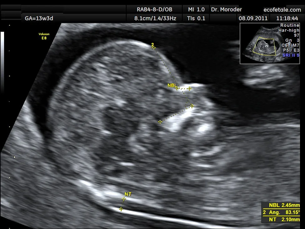
1ST TRIMESTER- NUCHAL TRANSLUCENCY
Book an appointment
Discuss your case with the physician and find out what the next steps are.
This scan is performed from 11 weeks and 1 day to 13 weeks and six days (CRL 45mm – 84mm). It is usually carried out transabdominally, but in some cases the examination may need to be done transvaginally.
Aims of the scan
The exact gestational age is established: This is particularly important for women who cannot remember the date of their last period, who have irregular periods, or who conceived while breastfeeding or soon after stopping the contraceptive pill. We measure the size of the fetus and from this calculate the expected due date (40 weeks).
Assessment of the risk of Down syndrome and other chromosomal abnormalities: Every woman should receive an assessment of her individual risk for this pregnancy. This is calculated by taking into account the mother's age, the measurement of two hormones in her blood (PAPP-A and free beta-hCG) and 4 ultrasound markers, which are: nuchal translucency (the fluid behind the back of the fetus's neck), which is the most important marker; the nasal bone; blood flow across the right side of the fetal heart (through the tricuspid valve); and blood flow in the fetal ductus venosus (a small vessel in the liver). Parents will receive full counselling about the significance of this risk and the various options for further testing.
Diagnosis of multiple pregnancy: About 2% of natural conceptions and 10% of IVF conceptions result in a multiple pregnancy. The scan can determine whether both fetuses are developing normally and whether the babies share the same placenta, which can lead to problems in the pregnancy. In such cases, it would be advisable to monitor the pregnancy more closely.
Diagnosis of certain major congenital anomalies: Major anomalies may be visible at this gestational age, but the 20-week scan is also essential.
Diagnosis of early pregnancy failure (missed miscarriage): Sadly, in 2% of women attending for the nuchal translucency scan, it is found that the fetus has died, often several weeks earlier and without any warning. Couples will receive full counselling about the possible causes of this problem and the options for any further steps that may be necessary.
Assessment of the risk of pre-eclampsia and fetal growth restriction: At the end of the examination, blood flow in the mother's uterine arteries will be measured in all patients by Doppler ultrasound. The result of this test can tell us which women are at increased risk of developing pre-eclampsia, mainly before 34 weeks, as well as which fetuses are at risk of growth restriction, mainly in the third trimester.
Assessment of the risk of preterm birth: At the end of the examination, the length of the cervix will be measured transvaginally in all patients who wish it. We know that during pregnancy the cervix should be long and closed. In women found to have a short cervix on examination, the risk of preterm birth is increased. Management in this case depends on the problem.
Individual risk of Down syndrome
The vast majority of babies are healthy. However, all women, regardless of their age, have a small risk of having a baby with a physical and/or intellectual disability. In some cases the abnormality is due to a chromosomal abnormality such as Down syndrome.
The only way to know for certain whether the fetus has a chromosomal abnormality is through an invasive diagnostic test, such as chorionic villus sampling (CVS) or amniocentesis. However, these tests carry a risk of miscarriage of around 1%.
It is up to you and your partner to decide whether or not the risk of the fetus having chromosomal abnormalities is high enough to justify an invasive test. As a guideline, an invasive test is usually offered if the risk of Down syndrome is 1 in 300 or higher.
The most accurate method of assessing the risk of the fetus having Down syndrome is carried out at 11-14 weeks of pregnancy and depends on the following:
- The mother's age
- The amount of fluid behind the back of the fetus's neck (nuchal translucency)
- The presence or absence of the fetal nasal bone
- The fetal heart rate
- Blood flow across the tricuspid valve of the fetal heart
- Blood flow through the ductus venosus in the fetal liver
- The presence or absence of any structural abnormalities
- The level of two hormones (free beta-hCG and PAPP-A) in the mother's blood
After the scan, the estimated risk of Down syndrome, based on all of the above factors, will be discussed with you. Only you can decide whether you want to undergo an invasive diagnostic test. Whether or not you decide to have an invasive test, a detailed scan at 20 weeks is recommended to check the fetus for structural abnormalities.
Related articles
- TestsGUIDE TO FIRST TRIMESTER ORAL TRANSPARENCY
- Short postsFIRST-TRIMESTER SCAN - NUCHAL TRANSLUCENCY
- Informative ArticlesPREGNANCY TEST FOR DOWN SYNDROME


He supports scientific excellence in clinical practice and recognizes the possibilities offered by modern reproductive methods to couples who wish to have their own child.
His profileThis information is for general guidance only and does not replace a medical examination and advice.